50% Response rate
China has become an important sourcing base for Medical Ultrasound Transducer products. Its manufacturers serve hospitals, diagnostic centers, veterinary clinics, and medical equipment brands worldwide. This introduction explores the companies behind these devices and the capabilities that distinguish them.
A reliable manufacturer must demonstrate more than competitive pricing. Buyers should examine probe frequency ranges, connector compatibility, cable durability, housing design, and sterilization requirements. A linear probe may support vascular imaging, while a convex probe often suits abdominal examinations. Small design details matter. A loose connector can interrupt a busy scanning session. A stiff cable can reduce clinical comfort.
This overview focuses on manufacturers with practical engineering experience, documented quality systems, and responsive technical support. Evidence may include ISO 13485 certification, product testing records, clear specifications, and verified customer applications. Regulatory documentation should also match the destination market. Claims deserve careful checking. No supplier list is perfect, and online information can become outdated. That limitation deserves attention.
The discussion considers OEM and private-label services, customization options, production capacity, and after-sales support. It also examines how suppliers manage transducer testing, image consistency, acoustic performance, and long-term reliability. These factors help purchasers compare factories more objectively. A factory visit or independent sample evaluation can reveal issues that brochures hide. In medical imaging, consistency matters. One attractive sample proves little.
The following sections introduce leading Chinese manufacturers and explain how buyers can assess them with professional judgment. The goal is not to promote one company blindly. It is to support safer, clearer, and more informed sourcing decisions.
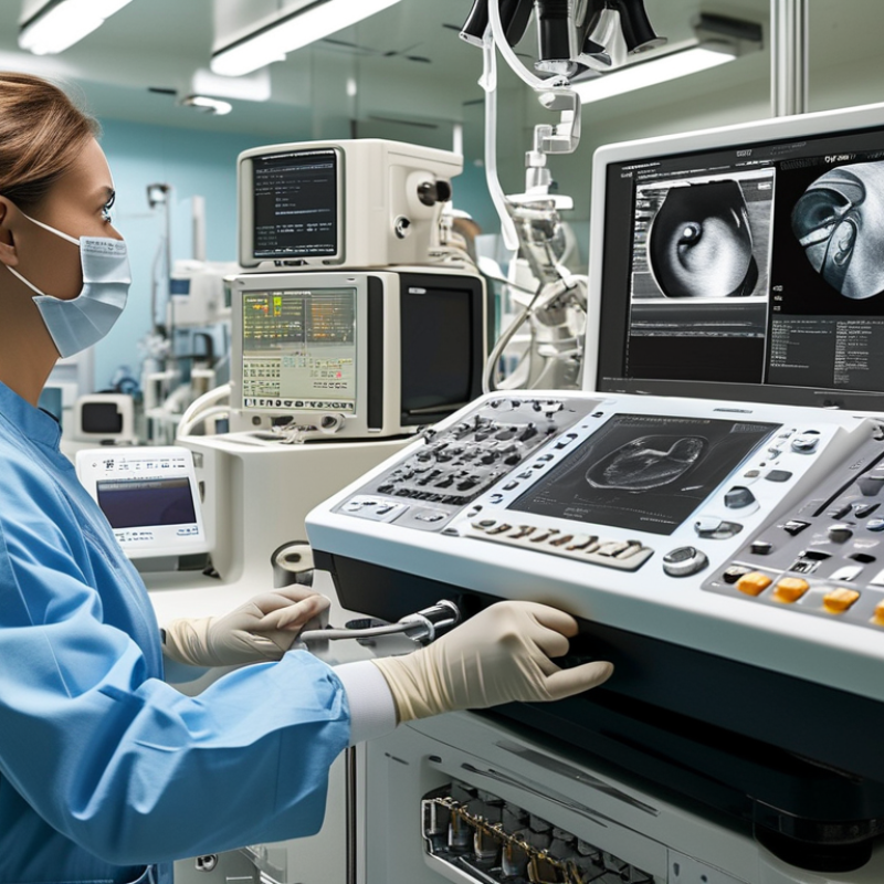
China’s medical ultrasound transducer manufacturers increasingly target probes covering 1–15 MHz. Lower frequencies support deeper abdominal imaging. Higher frequencies reveal finer details near the skin. Frequency alone, however, does not define clinical performance.
Grand View Research reported that the global ultrasound devices market was valued at approximately USD 8.9 billion in 2023. Its forecast indicates continued growth through 2030. MarketsandMarkets also projects steady expansion, driven by portable systems and point-of-care imaging. These figures suggest stronger demand for reliable transducers, not merely lower production costs.
IEC 60601-2-37 sets particular safety requirements for ultrasonic diagnostic and monitoring equipment. Manufacturers should verify acoustic output, thermal effects, mechanical effects, electrical insulation, and essential performance. Testing normally works with IEC 60601-1 and relevant electromagnetic compatibility requirements. Array uniformity matters in daily use. So do cable flexibility, connector stability, waterproofing, and cleaning resistance. A 1–15 MHz probe should maintain predictable sensitivity across its operating range. That sounds simple. It is not.
Experienced buyers should request test records, calibration methods, life-cycle data, and traceable quality controls. ISO 13485-based processes can strengthen manufacturing reliability, but certification alone cannot guarantee imaging quality. Clinical validation under realistic scanning conditions remains essential. Some market reports combine complete ultrasound systems with transducers, so their revenue estimates may overstate the probe segment. That limitation deserves attention.
China’s medical ultrasound transducer sector is expanding with global demand.
Fortune Business Insights valued the global ultrasound market at about USD 8.04 billion in 2023.
It projects a 6.1% compound annual growth rate through 2032.
Market growth alone does not prove manufacturing quality. Buyers should inspect evidence.
For China-based manufacturers, NMPA registration confirms approval for sale in China. It should match the exact transducer model, intended use, and classification. ISO 13485 certification shows a controlled medical-device quality system. It does not replace product registration.
For overseas sales, CE evidence should reflect Regulation (EU) 2017/745, including conformity assessment, technical documentation, risk management, and post-market monitoring.
Reviewers should also request IEC 60601-1, IEC 60601-1-2, and IEC 60601-2-37 test reports. Acoustic output, probe durability, sterilization compatibility, and biocompatibility need specific records.
A certificate alone can still hide gaps. This is where many evaluations become rushed.
Tips:
Ask for current certificates, registration numbers, model lists, and issuing-body details. Confirm that the legal manufacturer matches every document.
Check sample serial numbers and calibration records. Grand View Research reported steady growth in diagnostic ultrasound demand, but reports rarely measure probe reliability directly.
That limitation matters. In my experience, a clear traceability file is more useful than a polished brochure.
Some documents may look complete, yet revision dates or model names can conflict. Flag those details before clinical testing or procurement.
China’s medical ultrasound transducer manufacturers serve a broad clinical market, from routine abdominal scanning to specialized imaging. Grand View Research estimated the global medical ultrasound market above USD 8 billion in 2023, with steady growth through 2030. This demand is increasing interest in reliable probe categories, not only complete ultrasound systems.
2D probes remain common for general diagnosis and obstetric examinations. Convex probes provide deeper abdominal coverage, while linear probes show sharper detail in vessels, thyroid tissue, and superficial structures. Doppler transducers measure blood-flow direction and velocity. Small changes in crystal design can affect sensitivity. It is easy to overlook this during procurement.
3D and 4D probes support volume imaging, especially in obstetrics and selected surgical applications. Endocavity probes require careful ergonomic design, stable acoustic performance, and validated cleaning compatibility. MarketsandMarkets’ ultrasound equipment analysis identifies rising point-of-care use as a major growth factor, although forecasts vary by research method. Buyers should therefore review frequency range, bandwidth, connector durability, waterproofing, and clinical test records. Manufacturing experience matters. So does traceability. A lower quoted price may hide higher replacement costs, limited calibration support, or inconsistent image quality between batches. Personnel evaluating Chinese suppliers should request sample scans, quality certificates, lifecycle testing, and clear after-sales procedures before approval.
Technical comparison of commonly supplied medical ultrasound transducer categories
| Probe Category | Typical Frequency Range | Scanning Format | Common Clinical Applications | Typical Imaging Modes | Key Advantages | Main Considerations |
|---|---|---|---|---|---|---|
| 2D / B-Mode Probe | Approximately 2–15 MHz, depending on probe geometry and examination depth | Two-dimensional real-time imaging | General abdominal, obstetric, gynecological, musculoskeletal, vascular, and point-of-care examinations | B-mode; often available with color and spectral Doppler when supported by the probe and system | Versatile, widely used, and suitable for routine anatomical assessment | Image quality depends on frequency, aperture, acoustic coupling, and penetration requirements |
| 3D / 4D Volume Probe | Typically about 2–9 MHz for abdominal, obstetric, and gynecological volume imaging | Volumetric acquisition; 4D provides real-time or near-real-time volume imaging | Fetal face and anatomy, fetal cardiac assessment, pelvic imaging, gynecology, and selected abdominal studies | 2D, 3D, 4D, color Doppler, and volume-rendered imaging, depending on system capabilities | Provides multiplanar views and spatial anatomical information from a captured volume | Requires appropriate volume acquisition, patient cooperation, adequate acoustic windows, and compatible software |
| Doppler Probe | Approximately 2–10 MHz; continuous-wave probes may use dedicated lower-frequency elements | Directional flow and velocity assessment; may be integrated with 2D imaging | Peripheral vascular studies, carotid assessment, obstetric fetal heart-rate checks, cardiac examinations, and blood-flow evaluation | Color Doppler, power Doppler, pulsed-wave Doppler, and continuous-wave Doppler where supported | Assesses flow direction, relative velocity, vascularity, and hemodynamic patterns | Angle, sample-volume placement, aliasing, wall-filter settings, and scale selection affect measurements |
| Linear Array Probe | Approximately 5–18 MHz, with high-frequency models used for superficial structures | Rectangular or trapezoidal field of view | Thyroid, breast, tendons, muscles, nerves, small parts, superficial vessels, and ultrasound-guided procedures | B-mode, color Doppler, power Doppler, elastography, and needle-enhancement functions on compatible systems | High spatial resolution and excellent near-field detail | Higher frequencies generally provide less penetration; the relatively narrow footprint may limit coverage of deep or broad anatomy |
| Convex / Curvilinear Probe | Approximately 1–7 MHz for deeper abdominal and obstetric examinations | Curved, sector-like field of view with a broad far field | Abdomen, pelvis, obstetrics, urology, emergency assessment, and general deep-organ imaging | B-mode, color Doppler, pulsed-wave Doppler, harmonic imaging, and selected 3D/4D applications | Good penetration and wide coverage for deep organs and large anatomical regions | Lower frequencies provide depth but less superficial resolution; rib, bowel gas, and body habitus can reduce image quality |
| Endocavity Probe | Approximately 4–12 MHz, depending on endovaginal, endorectal, or other specialized design | Compact endocavitary field of view; commonly curved or sector-shaped | Transvaginal pelvic imaging, early pregnancy assessment, follicular monitoring, prostate imaging, and selected endocavitary procedures | B-mode, color Doppler, spectral Doppler, 3D volume imaging, and elastography on compatible systems | Close proximity to target anatomy supports high-detail imaging with a compact footprint | Requires strict infection-control procedures, protective covers where applicable, approved high-level disinfection, and trained operators |
| Phased Array / Cardiac Probe | Approximately 1–5 MHz for adult cardiac and deeper thoracic examinations; higher-frequency options may be used in pediatrics | Narrow sector field of view with electronic beam steering | Adult and pediatric echocardiography, emergency cardiac assessment, lung imaging, and intercostal examinations | 2D, M-mode, color Doppler, pulsed-wave Doppler, and continuous-wave Doppler where available | Small footprint fits between ribs and supports imaging of moving cardiac structures | Lower frequency favors penetration, while sector geometry may provide less near-field detail than a linear probe |
A practical benchmark begins with element count.
Medical probes commonly use 64, 128, or 256 elements. More elements can improve aperture control and image detail. However, electronics, matching layers, and manufacturing consistency matter just as much.
A 256-element probe with uneven element output may perform worse than a reliable 128-element design. During supplier evaluation, request element uniformity data, not only a specification sheet.
Bandwidth affects tissue penetration and resolution.
A broad range, such as 2–5 MHz or 5–12 MHz, supports different clinical applications. Higher frequencies show finer structures but lose penetration in deeper tissue.
Sensitivity should be reviewed with noise levels, transmit voltage, and focal depth. A high sensitivity figure alone can mislead.
Test images should include cyst contrast, axial resolution, and performance through a tissue-mimicking phantom.
IPX7 protection means temporary immersion under defined test conditions, commonly up to one meter for thirty minutes.
It does not prove resistance to disinfectants, repeated cable bending, or long-term fluid exposure.
Ask manufacturers for inspection records, sealing methods, and validation results based on IEC 60529.
We once focused too heavily on bandwidth and missed connector fatigue. That was a useful correction.
Real reliability appears during cleaning cycles, drops, thermal changes, and daily clinical handling. Manufacturing traceability also matters, including element inspection, calibration records, and final acoustic testing.
Export readiness depends on evidence, not factory claims. ISO 10993-1:2018 requires a risk-based biological evaluation for patient-contacting materials. For ultrasound transducers, teams should document contact duration, skin or mucosal exposure, and sterilization effects. Typical evidence includes cytotoxicity, sensitization, irritation, and chemical characterization tests. The FDA’s 2023 guidance on ISO 10993-1 warns against using a routine checklist without considering material and clinical risks.
Small details matter. A probe cable may pass before disinfection, then fail after repeated cleaning cycles. Residual adhesive can also change chemical results. This is easy to miss. Export files should link each material to supplier declarations, test samples, laboratories, and final production parts. Some Chinese manufacturers provide these records early. Others provide only summary certificates, which may not satisfy a cautious reviewer.
IEC 60601 testing adds electrical and functional evidence. Under IEC 60601-1, a commonly used Type BF applied part is assessed against patient leakage limits of 100 microamperes in normal condition and 500 microamperes under single fault conditions. IEC 60601-2-37 also addresses diagnostic ultrasonic equipment, including acoustic output and display-related safety controls. The FDA’s 2019 guidance on diagnostic ultrasound systems emphasizes thermal and mechanical index reporting. A credible data package should show test conditions, equipment calibration, software version, and retest results. One weakness remains: a passing laboratory report cannot prove every field scenario. Real cleaning, cable bending, and operator handling still deserve review.